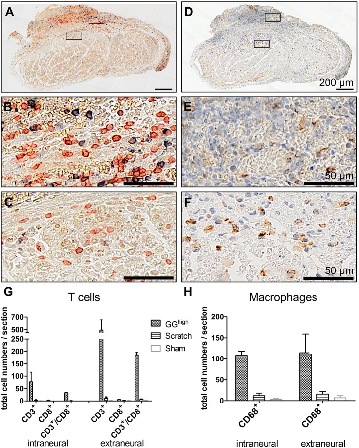
Fig. 4.
T-cell and macrophage recruitment at the sciatic nerve 3 weeks after glutaraldehyde glue application. (A-C) Cells were co-stained with antibodies against CD3 (red) and CD8 (blue) to detect T cells and (D-F) against CD68 (brown) for macrophages. Nuclei in D-F were counterstained with hematoxylin (blue). Quantification of cell numbers of representative cross sections revealed (G) high amounts of T cells and (H) macrophages inside and surrounding the sciatic nerve of glutaraldehyde-glue-treated rats in comparison to scratch and sham groups. Displayed are mean cell numbers with s.e.m. of respective histological sections of two rats of each experimental group.

