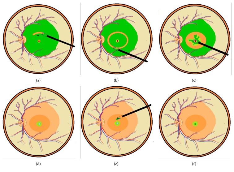
Figure 1.
Direct forceps grasping was used to create an ILM break where a good ICG staining had been obtained (a). A ring-shaped ILM flap was created around the macular hole (b). The ILM was detached from the retina to the edge of the macular hole (c). Further anterior ILM peeling was performed up to the arcade along with the overlying ERM. The ILM flap anchoring on the hole edge was inverted and inserted into the hole using microforceps (d). A small amount of Viscoat was then carefully applied on top of the hole. After the infusion was turned off, a piece of the previously obtained free ILM flap was released from the microforceps on top of the macular hole (e). The microforceps with closed tips was used to guide the ILM tissue to fall on the hole and to nudge the free ILM tissue into the hole (f).

