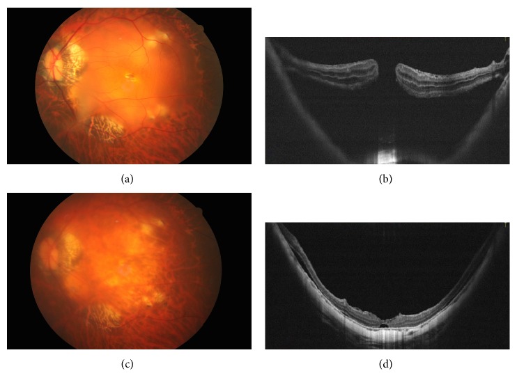
Figure 2.
A 62-year-old woman developed a macular hole with retinal detachment in the left eye. Preoperative fundus photograph and optical coherence tomographic image showed the macular hole with localized posterior retinal detachment and adherent epiretinal membrane (a, b). Fundus photograph and optical coherence tomographic images 2 months after surgery showed the sealed macular hole with shallow residual subretinal fluid (c, d).

