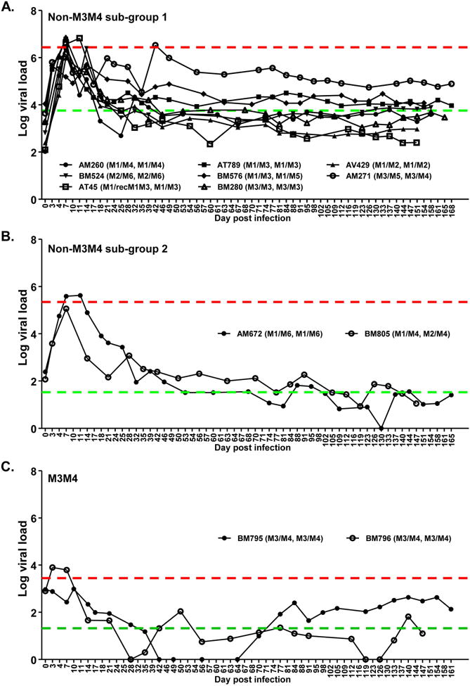
Figure 1. Viral load profiles of individual SIV-infected macaques.

Twelve female Mauritian cynomolgus macaques were vaginally infected with SIVmac251 and monitored for viral load (Log10 SIV RNA copy number/ml plasma) over time. Animal ID (MHC I haplotype, MHC II haplotype) is shown for each individual monkey. Red line: mean peak viral load of monkey group. Green line: Mean set point viral load of monkey group. (A) Non-M3M4 sub-group 1: Monkeys of non-M3M4 genotypes with poor SIV control. (B) Non-M3M4 sub-group 2: Monkeys of non-M3M4 genotypes showing SIV control. (C) M3M4: Monkey group of M3M4 genotype. Note that two more monkeys (with M3M4 and non-M3M4 genotypes, respectively) were challenged in the same experiments, but did not show detectable viral load and were excluded from study (See Table S1).
