
Figure 2. Effects of perfusion on bone formation in vitro.
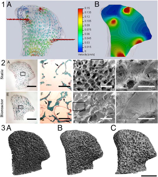
(1) Computational models of medium flow through TMJ constructs during bioreactor cultivation. (1A) Color-coded velocity vectors indicate the magnitude and direction of flow through the entire construct based on experimentally measured parameters. (1B) Construct is digitally sectioned, and the color-coded contours are used to indicate the magnitude of flow in the inner regions. (2A–H) Bone formation was markedly enhanced by perfusion, in a manner dependent on the fluid flow pattern. (2A–D) Constructs cultured under static conditions. (2E–H) Constructs cultured with medium perfusion. (2A, E) Trichrome staining of the entire cross-section of scaffolds showing differences in the new matrix distribution (red) compared with the original scaffold (green) for the static (2A) and perfused (2E) culture groups. (2B, F) Major differences in osteoid formation (arrows) in the central regions of constructs cultured statically (2B) and in perfusion (2F). (2C, D, G, H) SEM images of the central construct regions. (2C, D) Statically cultured constructs exhibit empty pore spaces and loosely packed cells. (2G, H) Constructs cultured in perfusion demonstrate the formation of dense and confluent lamellae of bone tissue that fill entire pore spaces. (Scale bars - 2A, E: 5 mm; 2B, C, F, G: 1 mm; 2D, H: 500 μm.) (3A–C) Architecture of the mineralized bone matrix developed over time and in a manner dependent on culture conditions. The reconstructions of 3D μCT images demonstrate the changes in pore structure (relative to the initial state) that were evident at the end of the 5th week of cultivation. (Scale bar: 5 mm.) Images reproduced with permission from reference (48)