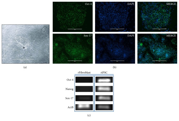
Figure 1.
Characterization of induced pluripotent stem cells (iPSs). (a) Morphology of the iPS cell colonies. (b) Immunofluorescent staining of rat-induced pluripotent stem cells for the expression of the pluripotency markers OCT-4 (upper panels, scale bar 400 μm) and SOX-17 (lower panels, scale bar 1000 μm) and overlay with the control staining of the nucleus with DAPI (Sigma). (c) RT-PCR analysis of gene expression in undifferentiated riPS cells.

