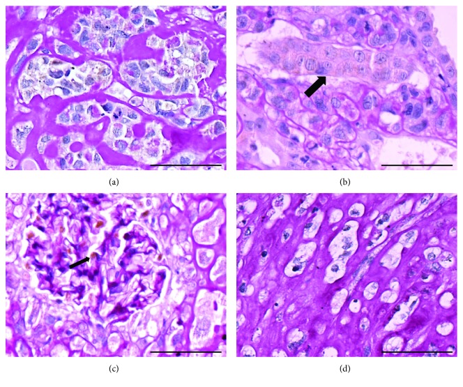
Figure 5.
(a) Nests of tumor composed with anaplastic cells with a light, heterogeneous cytoplasmic staining for WT1 antibody. WT1 immunohistochemistry revealed with diaminobenzidine (DAB) and followed by periodic acid-Schiff (PAS) staining. Bar: 50 μm. (b) Tumor cells surrounding a renal tubule. Notice the more homogeneous WT1 staining of the cytoplasm of the renal tubule (→) contrasting with the lightly reactive tumor cells. WT1 immunoperoxidase reaction revealed with DAB and followed by PAS staining. Bar: 50 μm. (c) Internal positive control of the reaction. Glomerular podocytes WT1 positive in histological section of chronic renal injured kidney treated with iPSs. Bar: 50 μm. (d) Negative control of the reaction. Histological section of an iPS-treated animal incubated with nonimmune rabbit immunoglobulin instead of the primary antibody. Bar: 50 μm.

