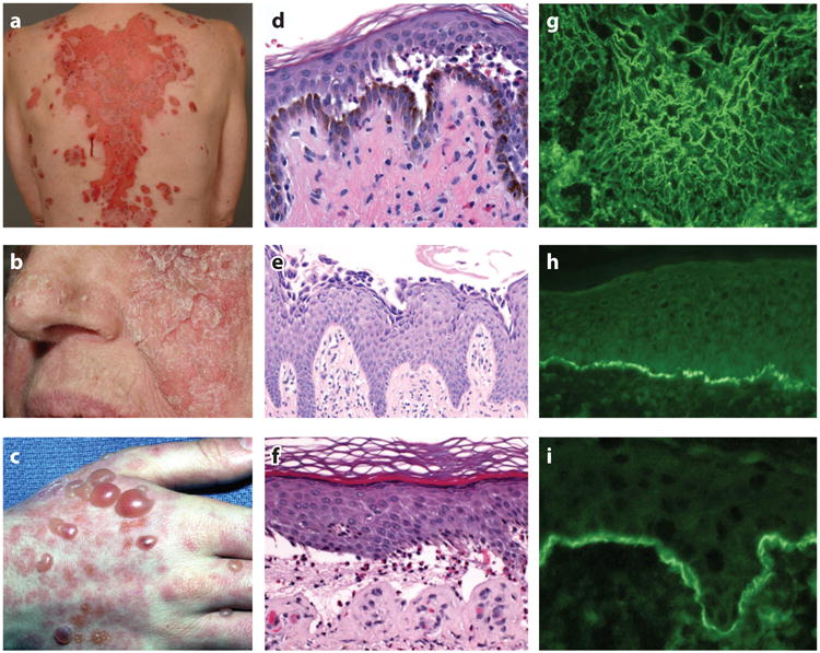
Figure 1.

Clinical, histological, and immunopathological findings in pemphigus and pemphigoid. Clinical images of (a) PV with large erosions, (b) PF with scaly crusted lesions, and (c) BP with tense blisters. (d) Histology of PV shows suprabasilar blister, (e) PF shows acantholysis in the granular layer of the epidermis, and (f) BP shows a subepidermal blister with eosinophils. Indirect immunofluorescence of (g) PV on monkey esophagus and (h) BP on normal human skin shows presence of circulating IgG binding cell surface and epidermal basement membrane, respectively. (i) Direct immunofluorescence of perilesional skin in a BP patient shows C3 at the epidermal basement membrane. Abbreviations: BP, bullous pemphigoid; C3, third component of complement; IgG, immunoglobulin G; PF, pemphigus foliaceus; PV, pemphigus vulgaris.
