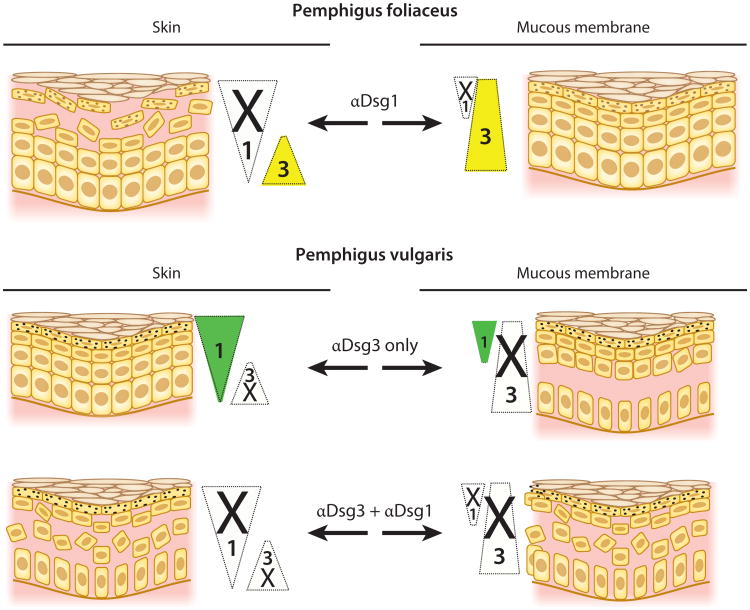
Figure 3.
Desmoglein compensation in pemphigus. Triangles show the usual localization of Dsg1 (green) and Dsg3 (yellow) in the epidermis (skin) and mucous membrane. Triangle width indicates the relative amount of Dsg present at each cell level. Loss of color in a triangle represents loss of function of that particular Dsg due to the presence of αDsg1 or αDsg3. In any area in which Dsg1 or Dsg3 function has been inactivated and the other Dsg is not present to compensate, a blister (shown as loss of cell-cell adhesion) occurs. Abbreviations: αDsg1, anti-Dsg1 antibodies; αDsg3, anti-Dsg3 antibodies; Dsg, desmoglein.

