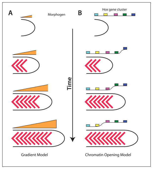
Figure 6. Models for Hox gene regulation.
Left, gradient model. As the embryo extends, the concentration of a secreted factor decreases, which provides a signal for the expression of more posterior Hox genes. It is also possible that a signal increases as the embryo extends. Right, Chromatin model. As the embryo extends, the chromatin opens up progressively in a 3′ to 5′ direction, allowing more posterior Hox genes to be expressed.

