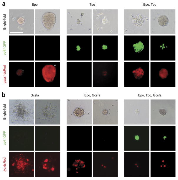
Figure 5.
Representative images of particular colonies grown in methylcellulose. (a,b) Progenitor cells isolated from fractionated WKM of adult (a) Tg(cd41:GFP, gata1:dsRed) and (b) Tg(cd41:GFP, lyz:dsRed) fish were grown for 4 d in the presence of zebrafish cytokines. (a) Epo stimulates growth and differentiation of small CFU-E (*) and large BFU-E (**) colonies that are hemoglobinized and express gata1:dsRed (left). Tpo stimulates growth and differentiation of relatively small CFU-T colonies that express high levels of cd41: GFP and low levels of gata1:dsRed (middle). Combinatorial addition of Epo and Tpo stimulates mixed CFU-TE colonies, consisting of clusters of erythrocytes and thrombocytes that express high levels of both cd41:GFP and gata1:dsRed (right). (b) Gcsf stimulates growth and differentiation of myeloid CFU-G/M colonies that express lyz:dsRed (left), whereas the combination of Epo and Gcsf encourages differentiation of hemoglobinized lyz:dsRed CFU-GEM colonies (middle). Combinatorial addition of Epo, Tpo, and Gcsf expands hemoglobinized CFU-GEMT colonies that express both cd41:GFP and lyz:dsRed (right). All photomicrographs were taken at original magnification ×200. Scale bar (top left) represents 100 μm in all images. Modified from ref. 17; originally published in Blood. Svoboda et al. Dissection of vertebrate hematopoiesis using zebrafish thrombopoietin. Blood. 2014;124:220–228. © The American Society of Hematology.

