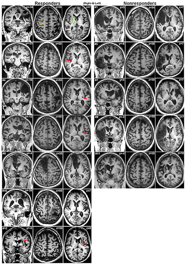
Fig. 3.
Magnetic resonance images for responders contrasted to nonresponders. For each subject, the left image shows the coronal slice at the level where the infarct is largest. The middle image shows a transverse slice at the level of the “hand knob” (Yousry et al. 1997) of primary motor area (M1), exemplified in subject #2 as yellow. The right image shows a lower transverse slice at the level of the internal capsule, exemplified in green. Not all transverse images show infarcts. Red arrows mark difficult-to-see infarcts. Responder status appears to be better indicated by posterior limb of internal capsule (PLIC) involvement than by M1 involvement. Slice z = 50 of subject #4 shows an infarct that largely affects M1, including some of the hand knob, yet this subject was a responder, likely because of only small involvement of the PLIC (slice z = 7). Conversely, nearly all the nonresponders (except subject #1) show no involvement of M1 at the level of the hand knob but substantial involvement of the PLIC (including subject #1).

