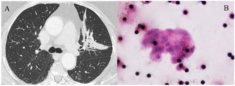
Figure 2.
(A-B): CTM in a case (number 63) of inconclusive FNA and core biopsy. (A) CT shows a consolidation in the left upper lobe with air bronchogram and small bronchiectases. (B) Filter (Hematoxylin and Eosin staining, original magnification x1,000) showing a CTM formed by more than 20 CTC with variably increased and irregularly shaped nuclei and multiple 8 µm pores of the filter appearing as black dots. Surgical pathology demonstrated a stage IIb adenocarcinoma.

