Figure 3.

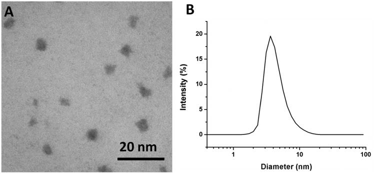
(A) Representative TEM image of hPR-DFO reveals ca. 4.0 nm spherical structures; (B) DLS size distribution of hPR-DFO dispersed in ddH2O displays a z-average diameter of ca. 3.5 nm with a PDI of 0.15.

(A) Representative TEM image of hPR-DFO reveals ca. 4.0 nm spherical structures; (B) DLS size distribution of hPR-DFO dispersed in ddH2O displays a z-average diameter of ca. 3.5 nm with a PDI of 0.15.