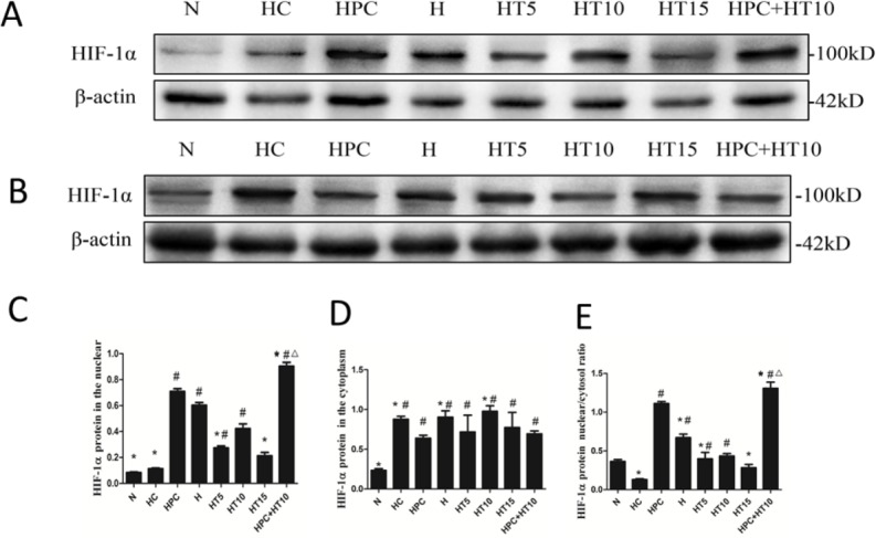
Figure 4. Effect of microtubule reticular changes on HIF-1 protein content.

(A) Expressions of HIF-1α in the nucleus. (B) Expressions of HIF-1α in the cytoplasm. (C) Relative protein levels for HIF-1α quantified in the nucleus. (D) Relative protein levels for HIF-1α quantified in the cytoplasm. (E) Ratio of nucleus HIF-1α to cytoplasm HIF-1α. The data showed that after hypoxia pre-treatment and/or taxol intervention, nucleus HIF-1α expression was increased while cytoplasmic HIF-1α expression was declined incardiomyocytes. After colchicine intervention, the nuclear/cytoplasmic ratio of HIF-1α was lower, suggesting that after colchicine injured microtubules, HIF-1α protein gathered in the cytoplasm instead of entering into the nucleus. #P < 0.05 vs. N group; *P < 0.05 vs. HPC group. △P < 0.05 vs. HT 10 group.
