
Figure 1. Chemical structure and characterization of Gal-G5.
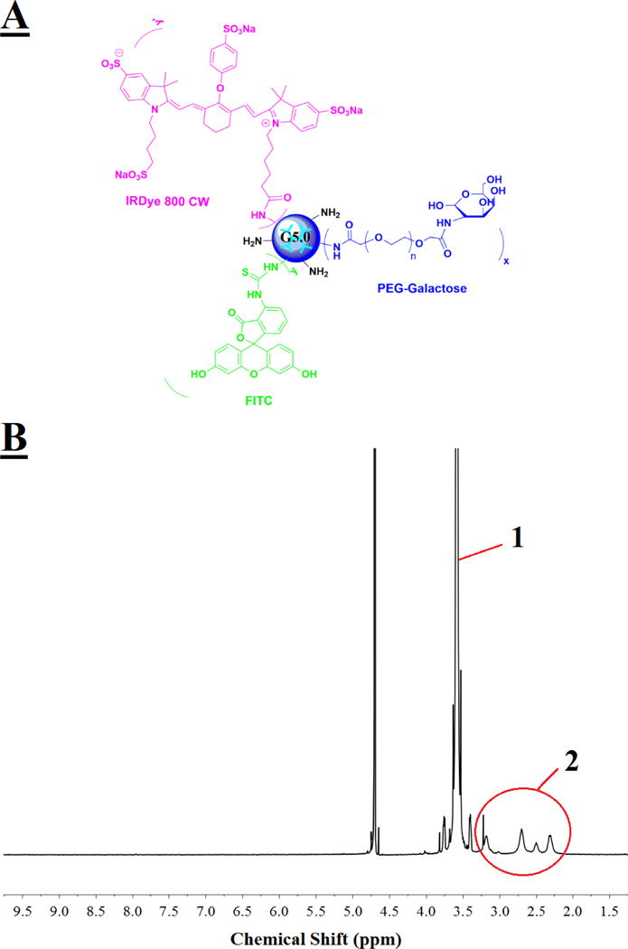
(A) PAMAM dendrimer G5 functionalized with hepatocyte specific ligand galactose alone or along with fluorescence dye (FITC or IRDye 800CW). (B) 300 MHz 1H NMR spectrum of Gal-G5. multiple proton peaks 2 between 2.2 and 3.4 ppm are assigned to the methylene protons of G5 branching units, while a singlet peak 1 at 3.6 ppm is assigned to the ethylene protons of the repeat units of PEG.