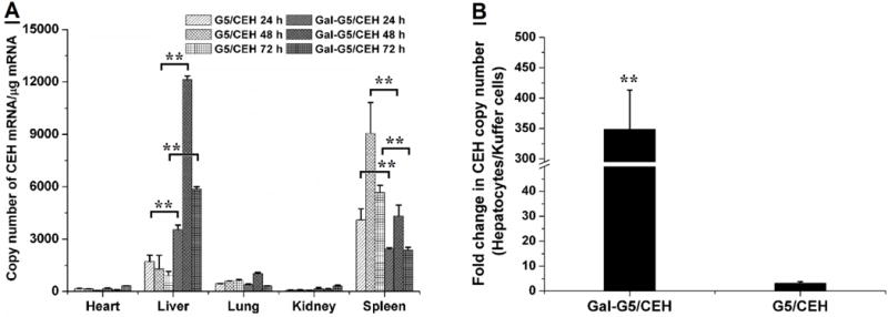
Figure 10.

Enhanced in vivo CEH expression in hepatocytes in the liver by Gal-G5/CEH. (A) CEH expression in the major organs at selected time points, i.e., 24, 48 and 72 h post-injection was quantified with RT-qPCR. n=3, **P<0.01. (B) In a parallel experiment, hepatocytes and Kupffer cells were isolated from the livers of the treated mice at 48 h-post injection, and CEH expression in the isolated cells was quantified with RT-qPCR. A significant increase in CEH expression in hepatocyte relative to that in Kupffer cells was achieved by Gal-G5/CEH. n=3, **P<0.01.
