
Figure 12. Increased CEH expression in the liver promotes RCT in vivo.
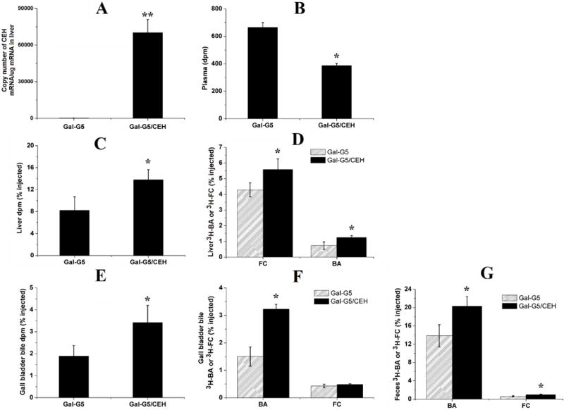
WT C57BL/6 mice were administered with Gal-G5 or Gal-G5/CEH via tail vein injection. After 48 h, mice were intravenously injected with [3H]-CE-labeled HDL. After an additional 48 h, blood, liver, gall bladder bile and feces were collected and analyzed. Radioactivity data (n = 3) are presented as the percentage of the total injected radiolabeled dose. (A) CEH expression in the liver (**P < 0.01); (B) radioactivity in plasma (*P < 0.05); (C) total radioactivity in the liver (*P < 0.05); (D) 3H-FC and 3H-BA in the liver (*P < 0.05); (E) total radioactivity in gall bladder bile (*P < 0.05); (F) 3H-FC and 3H-BA in gall bladder bile (*P < 0.05); (G) 3H-FC and 3H-BA in feces (*P < 0.05).