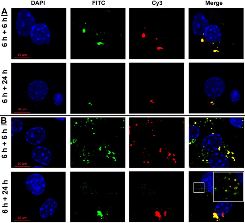
Figure 7. Intracellular dissociation of Cy3 plasmid from G5 or Gal-G5 in mouse hepatocytes.

Primary mouse hepatocytes were incubated with FITC-G5/Cy3 plasmid (4:1, w/w) (A) or FITC-Gal-G5/Cy3 plasmid (20:1, w/w) (B) in William's E medium supplemented with 10 % FBS for 6 h. The medium was replaced with fresh medium/10 % FBS, and the hepatocytes were incubated for an additional 6 or 24 h. At the end of the incubation, the cells were rinsed with DPBS, and the nuclei stained with DAPI and imaged by using confocal microscopy (630×). Blue, DAPI; Green, FITC; Red, Cy3. A magnified view of the area outlined by the smaller squared box is presented in the top right corner to show dissociation of plasmid from the carrier.
