
Figure 2.
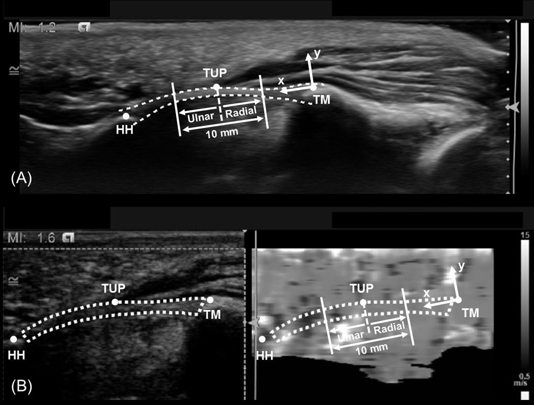
Representative B-mode (A) and ARFI-mode (B) ultrasound images that indicate the anatomically defined coordinate system, TCL (dotted line), hook of the hamate (HH), thenar muscles ulnar point (TUP), and ridge of the trapezium (TM). Additionally, the central 10 mm region of the TCL is identified in each image, along with the radial and ulnar regions used for analysis.