
Figure 6.
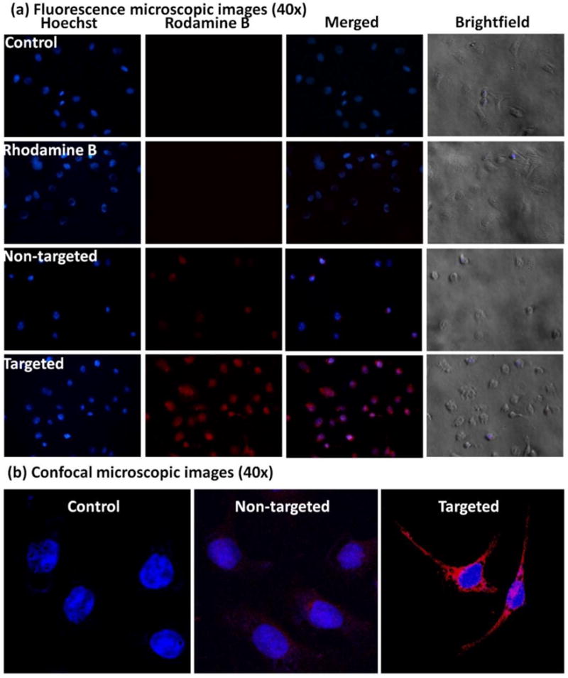
(a) Fluorescence microscopic images (40×) of SKOV3 cells incubated with nuclear stain Hoechst (blue fluorescence) and Rhodamine B (red fluorescence) labeled non-targeted and targeted formulations at 6 h are shown; (b) Confocal images of SKOV3 cells treated with DRAQ5® stain alone (control, blue fluorescence) or DRAQ5 and Rhodamine B (red fluorescence) labeled non-targeted formulation, and targeted formulation respectively are shown. Higher cellular uptake of the targeted formulation is suggested by the higher red fluorescent signal of Rhodamine B.