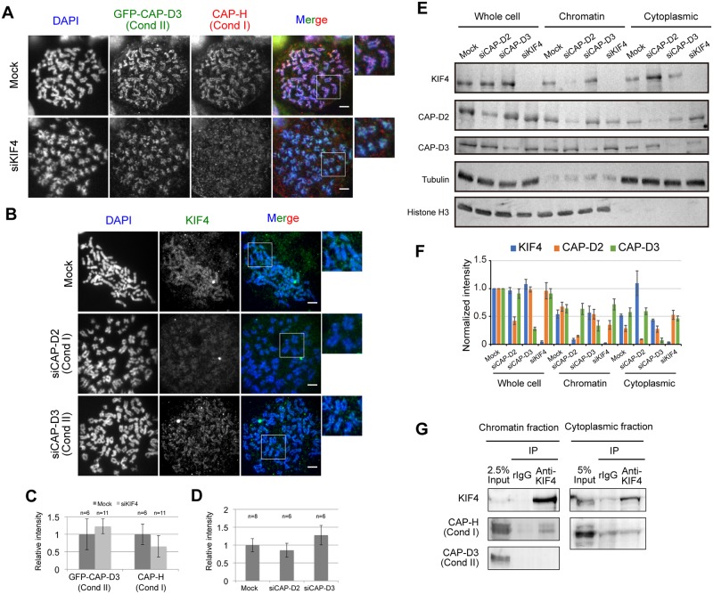
Fig 2. Interdependency between KIF4, condensin I and II.
(A) Localization of hCAP-H (condensin I) and GFP-hCAP-D3 (condensin II) in mock- or KIF4-depleted HeLa cells were confirmed by immunostaining. DNA was counter stained with DAPI. Scale bar, 5 μm. Close-ups of the regions indicated by white boxes were shown on the right. (B) Localization of KIF4 in mock, hCAP-D2-depleted or hCAP-D3-depleted HeLa cells were confirmed by immunostaining. DNA was counter stained with DAPI. Scale bar, 5 μm. Close-ups of the regions indicated by white boxes were shown on the right. (C) Quantification of relative mean fluorescence intensity of GFP-hCAP-D3 or hCAP-H staining in cells transfected with mock or KIF4-siRNA as indicated. Fluorescence intensities are normalized to the average pixel intensity of mock-transfected cells from the same experiment. Error bars represent the standard deviation. Two independent experiments were performed and data shown are from one representative experiment. (D) Quantification of relative mean fluorescence intensity of KIF4 staining in cells transfected with mock, hCAP-D2—or hCAP-D3-siRNA as indicated. Fluorescence intensities are normalized to the average pixel intensity of mock-transfected cells from the same experiment. Error bars represent the standard deviation. Two independent experiments were performed and data shown are from one representative experiment. (E) Western blot analysis of whole cell extract, chromatin fraction and cytoplasmic fraction prepared from mitotic mock, hCAP-D2-depleted, hCAP-D3-depleted or KIF4-depleted HeLa cells. Tubulin and histone H3 were used as loading controls. (F) The band intensity of (E) was quantified using ImageJ software and normalized against protein amount in whole cell extract of mock. At least three independent experiments were performed. (G) Physical interaction between KIF4, condensin I and condensin II. KIF4 was immunoprecipitated from chromatin-bound or chromatin-unbound protein fractions. Co-immunoprecipitation of hCAP-H (condensin I) and hCAP-D3 (condensin II) with KIF4 was analyzed by western blot. n = 3.

