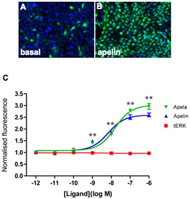
Fig 2. [Pyr1]apelin-13 and apela-32 activates ERK1/2 in CHO-B78 cells.
Cells were treated for 10min at 37°C with the indicated concentration of ligands. Representative thumbnail images acquired by the IN Cell Analyzer 1000 show immunohistochemical staining for intracellular ppERK1/2 (green) after treatment of CHO-B78 cells with vehicle (A) or 100nM [Pyr1]apelin-13 (B). The workstation can automatically demarcate nucleus from cytoplasm according to DAPI (blue) nuclear staining. A dose-response curve (whole-cell immunofluorescence expressed in arbitrary units) for [Pyr1]apelin-13 (blue) or apela-32 (green) is shown in (C). There are no measurable dose-dependent changes in tERK levels (red) in cells stimulated with apela-32—a similar result was obtained with cells stimulated with [Pyr1]apelin-13. The [Pyr1]apelin-13 and apela-32 stimulations were performed in separate cell wells (cells were used from the identical CHO-B78 passage number) and processed for ERK immunohistochemistry in the same experiment. Data are expressed as mean ± SEM, averaged from two separate experiments, each ligand concentration or vehicle (basal) with triplicate wells, and at least triplicate fields within wells. **p<0.01 comparing stimulations to basal conditions.

