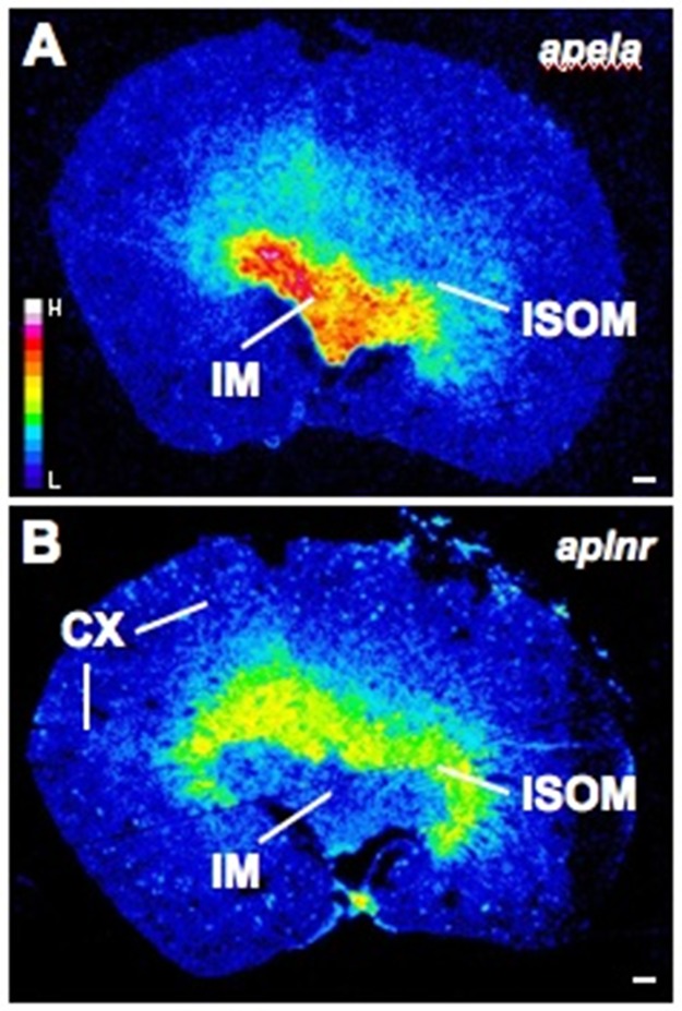
Fig 3. Representative autoradiographic film images demonstrating apela (A) and aplnr (B) expression in serial sections of a kidney from an adult male Wistar rat.
Apela is highly expressed in the inner medulla (IM) whereas aplnr labelling is primarily in the inner stripe of the outer medulla (ISOM). In contrast to the apela labelling, the aplnr probes also label a subpopulation of glomeruli (patchy ‘dots’) in the cortex (CX). The film images were exposed to film for 3 months, developed, scanned, exported to Image J and pseudocoloured. In these images yellow-red designates high expression whereas blue-black represents negligible or no labelling (see pseudocolour scale bar in (A)). The images are representative of results obtained from the kidneys of 8 rats. Scale bar = 500μm.

