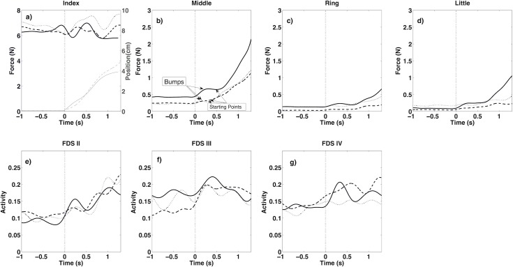
Fig 2.
Force (thick lines) and position (thin lines) of target finger, index (a), forces of non-instructed fingers (b-d) and EMGs of FDS muscle for related fingers (e-g) during static phase (Time: -1 to 0) and dynamic phase (Time: 0 to 1.35) (see the first paragraph of “Experimental protocol” section). Data presented in this figure were related to one representative subject during 3 trials (solid, dashed and dotted lines). Vertical dashed line indicates the start of movement.

