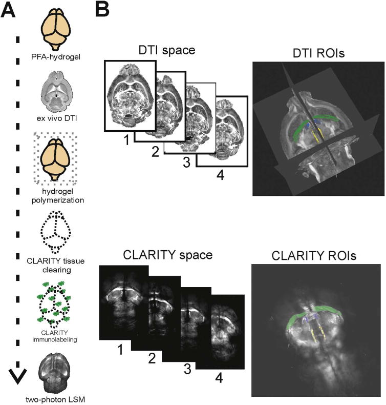
Fig. 1.

Brain-wide analyses combining ex vivo DTI and CLARITY. (A) Schematic of experimental design for DTI-CLARITY multimodal approach. (B) DTI images were registered to a single reference brain within DTI space. CLARITY MBP images were registered to the same reference brain within CLARITY space. WM ROIs were manually delineated in DTI FA space (TrackVis) and CLARITY MBP space (Imaris). Our procedure yielded corresponding WM ROIs that correlated in a near linear relationship between volume comparisons (Fig. 3B). Displayed ROI volumes are: hippocampal commissure (green), stria medullaris (blue), and fasciculus retroflexus (yellow). (For interpretation of the references to color in this figure legend, the reader is referred to the web version of this article.)
