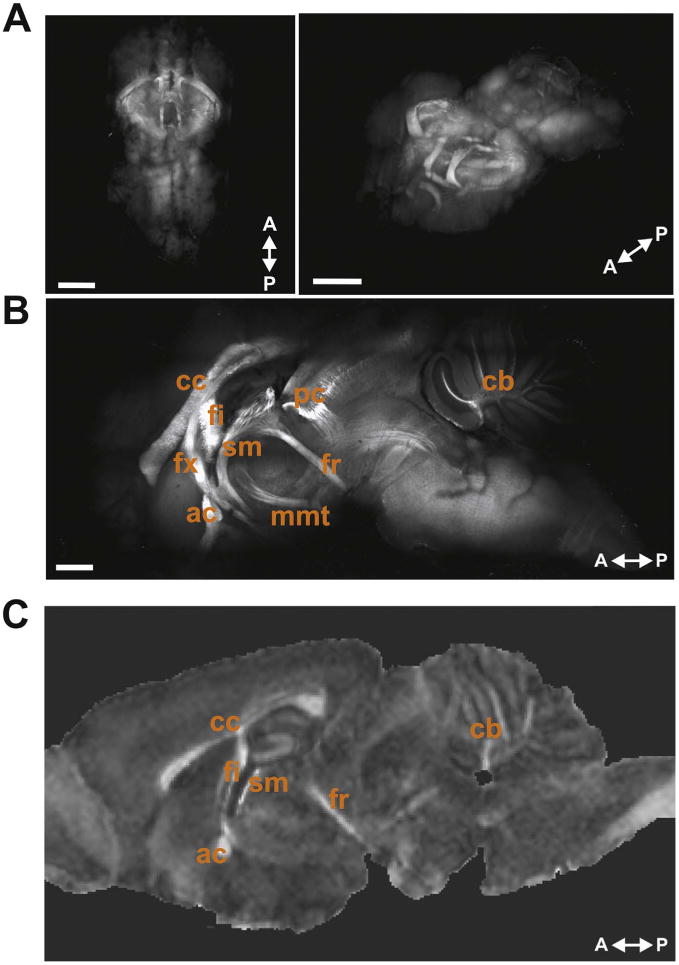
Fig. 2.

Whole mouse brain CLARITY MBP immunolabeling. (A) Images of CLARITY MBP whole brain in 3D showing a top-down view (left) and rotated horizontal view (right). Scale bars, 2 mm. (B) 1500 µm sagittal optical section near the midline of a CLARITY MBP brain shows many of the major myelinated WM structures within the mouse brain. Labeled WM tracts are: corpus callosum (cc), fimbria (fi), fornix (fx), anterior commissure (ac), stria medullaris (sm), posterior commissure (pc), fasciculus retroflexus (fr), mammillothalamic tract (mmt), and cerebellar WM (cb). Scale bar, 1.0 mm. (C) Representative sagittal image slice from FA map showing some of the same WM structures.
