Fig. 3.

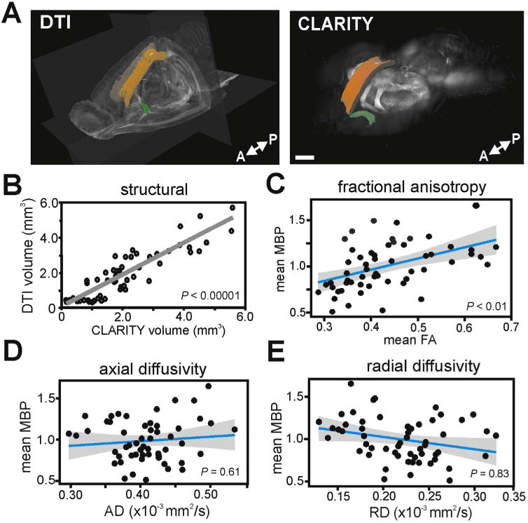
FA is correlated with MBP immunofluorescence. (A) Images show ROI-based comparisons of WM ROIs in DTI space (left) and MBP-positive WM structures in CLARITY space (right). Displayed ROIs are corpus collosum (genu, body, splenium) in orange and anterior commissure (posterior aspect) in green. Scale bar, 1000 µm. (B) DTI volumes significantly correlated with CLARITY volumes across all quantified WM structures (P<0.00001). (C) Mean MBP immunofluorescence correlated significantly with mean FA (P< 0.01). However, MBP did not correlate with measures of directional diffusivity, mean AD (D) or mean RD (E) across all WM ROIs measured. Linear regressions are shown with 95% confidence intervals. Each data point is a mean MBP immunofluorescence and mean FA measure from a single WM ROI, in one mouse brain. MBP immunofluorescence values were normalized to a global average. (For interpretation of the references to color in this figure legend, the reader is referred to the web version of this article.)
