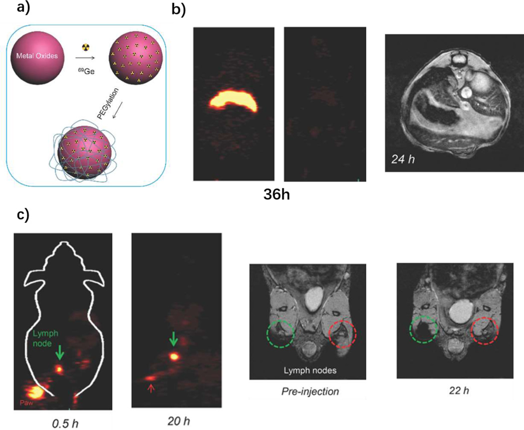
Figure 5.
69Ge-labled SPION for PET/MRI. a) Schematic figure of 69Ge labeling SPIONs; b) biodistribution monitoring post i.v. injection through PET and MRI. Contrast agents demonstrated long-term signals 36 h post injection with PET imaging and 24 h with MRI; c) Long-term lymph node mapping with PET and MRI. The green circle areas for MRI images are contrast agents injected position and the red areas are control areas. Reproduced with permission[90]. Copyright 2014, Wiley.

