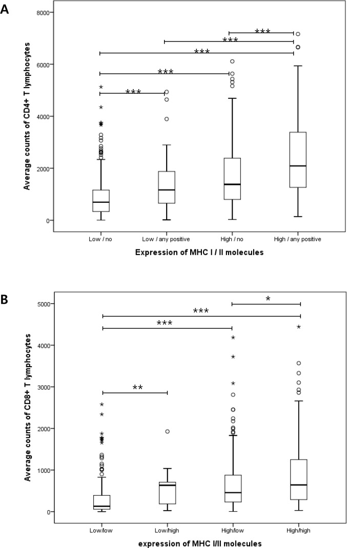
Fig 2.
The amount of CD4-positive (A) and CD8-positive (B) lymphocytes according to protein expression of MHC-I and -II in tumor cells. The patients were divided into four groups by combining MHC-I and -II expression levels. MHC-I was dichotomized by the mean value of its expression. In case of MHC-II, ‘no’ designates the cases with total negativity for MHC-II, and the others who have tumors with positivity for MHC-II regardless of percentage and intensity are designated as ‘any positive’. (*p < 0.05; **p < 0.01; ***p < 0.001).

