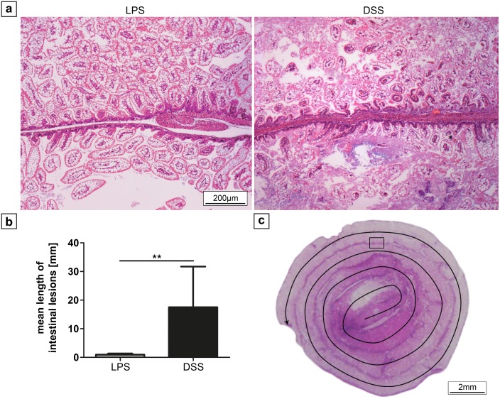
Fig 3. Histological changes of the small intestinal tissue after 72 h treatment.
(a) Representative ileal sections of neonatal mice from controls, the LPS, and DSS group (magnification 200x), showing healthy villi-structure versus injured intestinal segments (LPS: NEC score 2; DSS: NEC score 4) in the treatment groups. Severity of intestinal lesions and the incidence of NEC (b) as assessed by the NEC score was similar in both groups (n = 10). *** p < 0.001.

