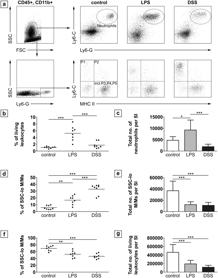
Fig 5. FACS analysis of leukocytes in the lamina propria of the small intestine of neonatal mice.
(a) Gated living CD45+, CD11b+ cells were either further gated on SSCint/hi, Ly6-G and Ly6-C double positive cells to identify neutrophils or on SSClo, Ly6-G-, Ly6-C and MHC II to identify intestinal monocytes/macrophages (M/M) populations (n = 8 per group). The percentage (b) as well as the total number (c) of neutrophils was significantly higher in the LPS group than in the DSS group and in controls, which did not differ among each other. Conversely, the percentage (d) of Ly6-Chi “inflammatory monocytes” (P1 gate) was higher in the DSS group as compared to the LPS group and controls. The total number of SSClo monocytes/macrophages was significantly lower in both treatment groups compared to controls. The percentage of resident macrophages found in the “incl. P3, P4, P5” gate (f) was significantly decreased after treatment compared to controls. (g) Total numbers of living leukocytes was significantly decreased in both treatment groups. * p < 0.05 ** p < 0.01, *** p < 0.001; SI = small intestine.

