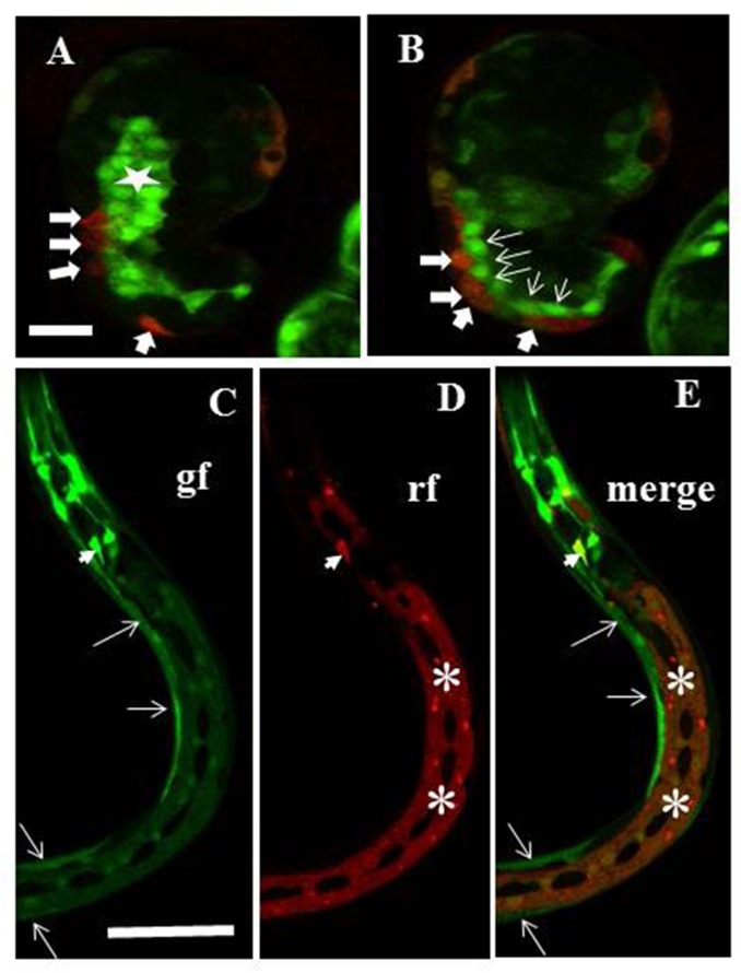
Fig 8. ngat-1p::gfp transcriptional reporter expression pattern.
A chromosomally integrated ngat-1p::gfp transcriptional reporter strain was made as described in Materials and methods. A dpy-7p::mCherry transcriptional reporter was injected into the ngat-1::gfp integrated line to create a non-integrated marker for epidermal cells (see Materials and methods), which may not mark all epidermal cells in a given animal because of potential mitotic loss of the extrachromosomal array. (A-B) merge of gfp and mCherry signals showing presumptive mCherry-expressing epidermal cells (thick arrows) that co-express gfp (as indicated by orange tinge) plus presumptive muscle cells (thin arrows), and presumptive intestinal cells (star) that express only ngat-1p::gfp in a comma stage embryo. (C-E) The ngat-1p::gfp reporter is expressed in body wall muscles (arrows in C and E) and in lateral epidermis (asterisks in D and E) in all post-embryonic larval stages. Some unidentified elongated cells in the head also express gfp and at least one (arrowhead) also expresses mCherry. (Dark oval-shaped areas in the lateral epidermis are lateral seam cells). rf = red fluorescence channel; gf = green fluorescence channel. Scale bar in panel A (for A and B) is 12.5 μm. Scale bar in panel C (for C-E) is 100 μm.

