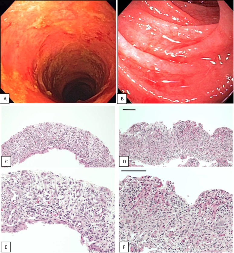
Fig 2. Endoscopic appearance (A, B) and histology (C-F) of denuded mucosa in the colon (left) and duodenum (right) at baseline in two patients who experienced CR of GVHD and survived more than one year.
Surface epithelium and crypts are virtually absent in the colon and duodenum and villi are absent in the duodenum (C,D H&E x10; E,F H&E x20; bar size = 100uM).

