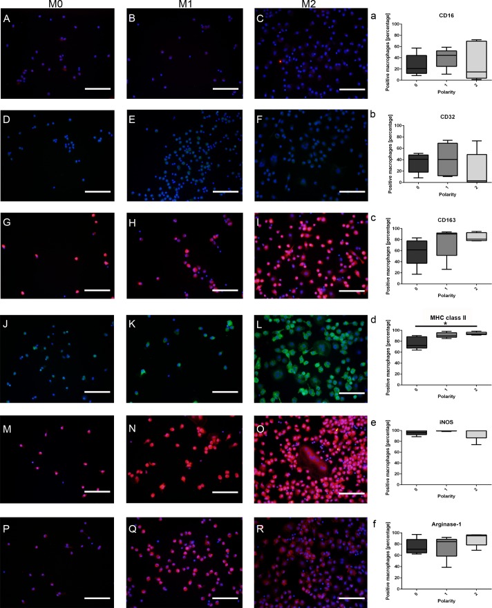
Fig 2. Immunofluorescence staining of in vitro cultured canine M0-, M1-, and M2-macrophages labeled with prototypic literature-based antibodies for the M1- (CD16, CD32, MHC class II, and iNOS) and M2-phenotype (CD163, CD206, and arginase 1), respectively.
A-C) Low to moderate membranous staining of M0-, M1-, and M2-macrophages for CD16. D-F) Likewise, CD32 shows a low to moderate staining in all three treatment conditions. G-I) M0-, M1-, and M2-macrophages demonstrate a moderate to high membranous staining with CD163. J-L) Intense membranous staining of M0-, M1-, and M2-macrophages with an anti-MHC class II antibody. M-O) Strong intracytoplasmic labeling of macrophages in all treatments (polarity 0, 1, 2) for inducible nitric oxide synthase (iNOS). P-R) High intracytoplasmic expression of arginase 1 in small/roundish, amoeboid, and spindeloid macrophages as well as in multinucleated giant cells (MNGs). a-f) Statistical evaluation of the mean expression percentages of prototypic M1-/M2-markers evaluated in 5 dogs and related to the polarization state of the macrophages (polarity 0, 1, 2). Note that, except for MHC class II, none of the remaining tested antigens (CD16, CD32, iNOS, CD163, and arginase-1) were differently expressed between canine M0-, M1-, and M2-macrophages (* = p≤0.05; Kruskal-Wallis-Test with pairwise Mann-Whitney-U-Tests). Scale bars = 100 μm. Nuclear counterstaining with bisbenzimide.

