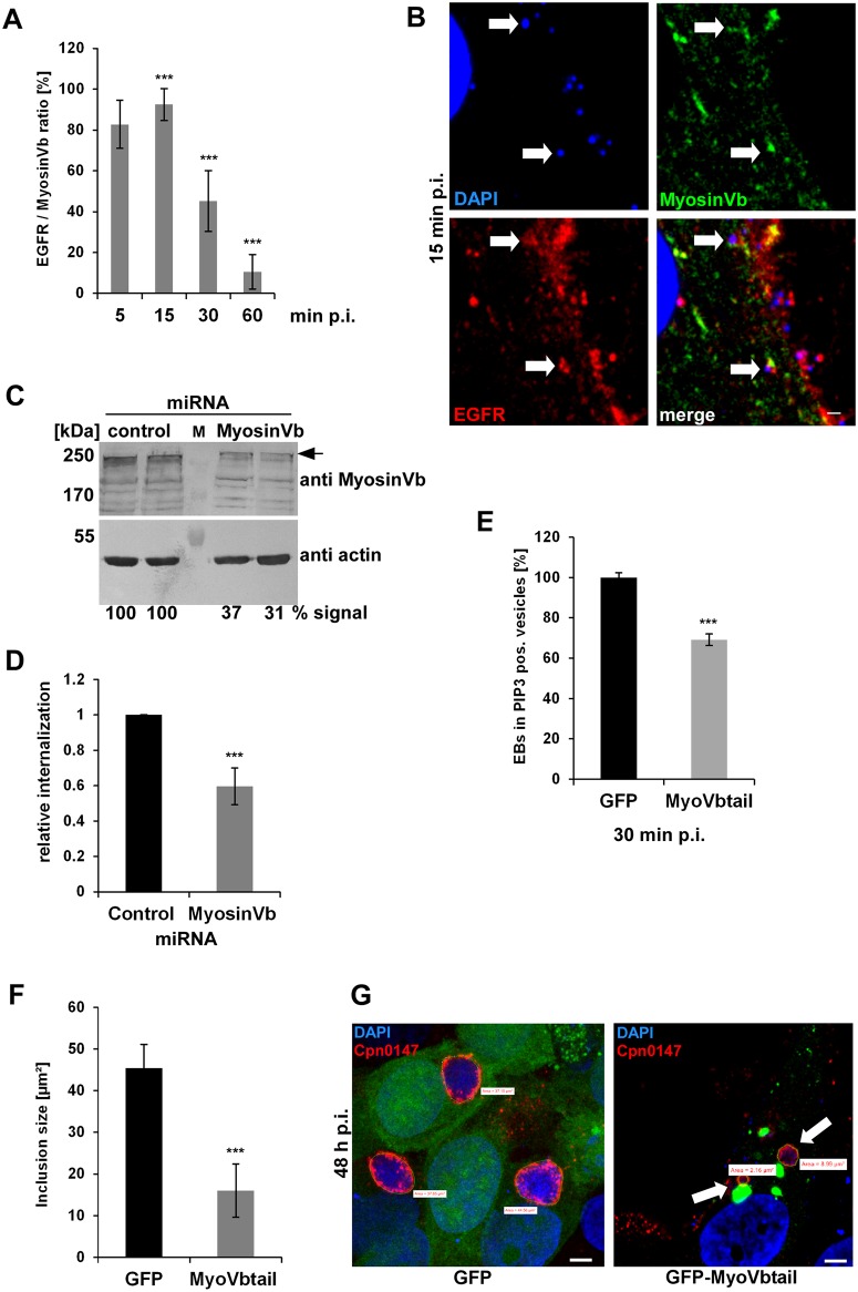
Fig 6. The acquisition of myosin Vb by the Rab11-Fip2 adaptor protein is essential for the C. pneumoniae infection.
(A) Quantification of colocalization of EGFR-positive EBs with GFP-MyosinVb from 5 min to 60 min p.i. (n = 3). (B) Confocal images of colocalization of GFP-MyosinVb and EGFR with C. pneumoniae EBs at 15 min p.i. White arrows indicate colocalization. Bar 1 μm. (C) Immunoblot analysis of cells transiently transfected for 72 h with control or myosin Vb miRNA plasmids and lysed in phospho-Lysis buffer. Samples were fractionated by SDS/PAGE and probed with antibodies against myosin Vb to monitor knockdown; β-actin served as loading control. The pixel intensity of bands was analyzed with ImageJ. The arrow marks the specific myosin Vb band. (D) Relative internalization of EBs into cells transfected for 72 h with control or MyosinVb miRNA plasmids was measured by q-PCR at 2 hpi as described in the legend to Fig 4 (n = 6). (E) Quantification of internalization of EBs into PI3P endosomes at 30 min p.i. Confocal images of 30 individual cells transiently expressing mCherry-2xFYVE and GFP or GFP-MyoVbtail were analyzed (n = 3). (F) Quantification of the inclusion diameter in cells expressing in GFP or GFP-MyoVbtail cells at 48 hpi. Inclusions were stained with anti-Cpn0147 and anti-rabbit Alexa594 and their diameters were measured in 30 individual cells (n = 3). *** P value ≤0.001, n.s. P value ≤0.01 (G) Confocal images of cells quantified in (F). Arrows mark the smaller inclusions in cells expressing GFP-MyoVbtail. Green lines mark the outline of the inclusions used for quantification. Bar 5 μm.

