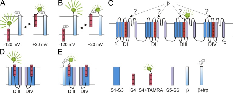
Figure 1.
Using VCF to identify conformational changes in NaV1.5. (A) A fluorophore attached to the voltage sensor S4 could experience changes in its microenvironment when S4 moves outward in response to a membrane depolarization. The change in microenvironment alters the fluorescence from the fluorophore, e.g., by changes in the hydrophobic/hydrophilic nature of the environment or by approaching a quenching residue. (B) Similarly, the fluorescence from a fluorophore attached to an immobile protein segment could change when S4 moves outward, if the outward moving S4 changes the microenvironment around the fluorophore. (C) Using VCF, one can label, one at a time, the four different S4s with a fluorophore (here shown the construct with the fluorophore attached to DIII-S4). When each of these constructs, one at a time, are coexpressed with β1 or β3, one can detect whether β1 and/or β3 alter the S4 movement in a specific domain (here DIII). (D and E) By introducing a quenching tryptophan residue in a β subunit (E), one can detect whether the β subunit is close to an S4 in a specific domain. If the β subunit is located close to DIII-S4, then one would expect to see a tryptophan-induced change in the fluorescence signal from the construct with a fluorophore attached to DIII-S4 (compare fluorescence in D and E).

