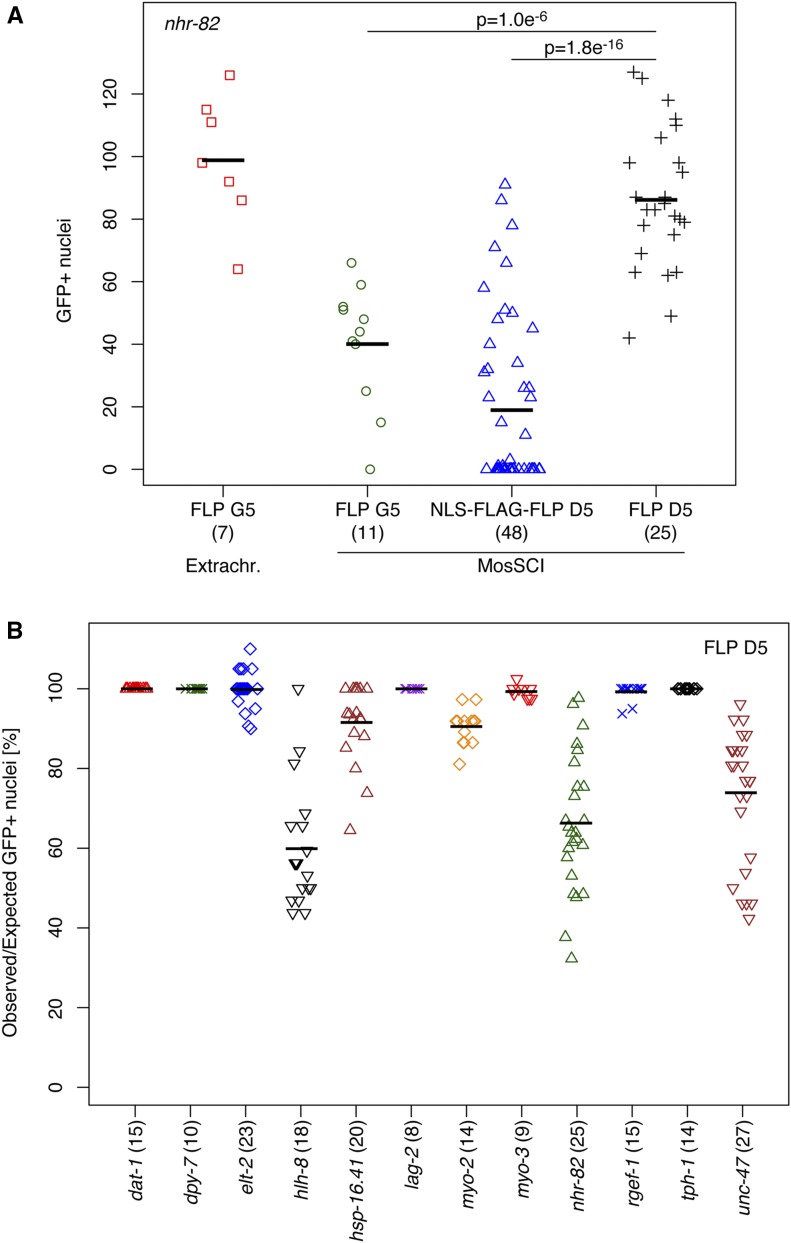
Figure 2.
Optimization and quantification of FLP activity. FLP activity was evaluated based on the number of nuclei expressing GFP::HIS-58 in animals carrying the integrated dual color reporter described in Figure 1. (A) Comparison of strains expressing FLP from an extrachromosomal array (FLP G5; red squares) or from integrated single-copy transgenes (MosSCI): FLP G5 (green circles), NLS-FLAG-FLP D5 (blue triangles), or FLP D5 (black crosses). The same nhr-82 promoter sequence was used in all strains. Shown are absolute numbers of GFP-positive nuclei; numbers in brackets indicate the number of adults evaluated for each strain; black horizontal lines represent the means. P values were obtained through unpaired, two-sided t-tests, indicating that FLP D5 has the highest activity among the MosSCI strains. (B) Comparison of MosSCI strains expressing FLP D5 under control of different cell type-specific or inducible promoters: dopaminergic neurons (dat-1; red triangles), hypodermis (dpy-7; green crosses), intestine (elt-2; blue diamonds), M lineage (hlh-8; black triangles), heat inducible (hsp-16.41; brown triangles), notch signaling (lag-2; purple crosses), pharynx (myo-2; orange diamonds), body wall muscles (myo-3; red triangles), seam cell lineage (nhr-82; green triangles), pan-neuronal (rgef-1; blue crosses), serotonin-producing neurons (tph-1; black diamonds), and GABAergic motor neurons (unc-47; brown triangles). Shown are the percentages of expected nuclei expressing GFP::HIS-58; numbers in brackets indicate the number of animals evaluated for each strain; black horizontal lines represent the means. For three promoters, on average, FLP activity was observed in 60–70% of the expected nuclei; for the remaining promoters, the mean activity was > 90% (see Table 1). GABA, γ-aminobutyric acid; MosSCI, Mos1-mediated single-copy insertion; NLS, nuclear localization signal.

