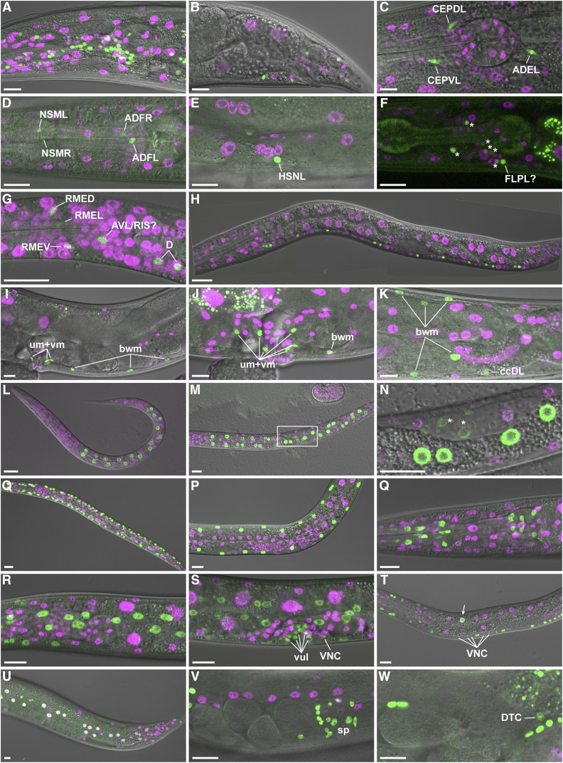
Figure 3.
Tissue-specific FLPase activity. Transgenic animals expressing FLP D5 in specific cell types and the dual color reporter were observed by live confocal microscopy. (A and B) Promoter of rgef-1 (Prgef-1); projection of eight confocal sections [(A) adult head lateral view] or single confocal section [(B) adult tail lateral view]. (C) Pdat-1; projection of four confocal sections of head of L4 larva. Note that the green channel represents coexpressed GFP::HIS-58 and mNeonGreen in this strain (see Figure S3). (D and E) Ptph-1; projection of three confocal sections [(D); adult head lateral view] or a single confocal section [(E); adult vulva lateral view]. (F) Pmec-7; projection of seven confocal sections of head of L4 larva. To ease visualization of the weak GFP signal, the corresponding DIC image was not included in the merge. Asterisks indicate six unidentified neurons, whereas a seventh neuron might be FLPL. (G and H) Punc-47; late L1; projection of four confocal sections [(G); head lateral view] or stitch of single confocal sections (H). (I–K) Phlh-8; single confocal section [(I); adult vulva lateral view] or max projections [(J); vulva region, 8 confocal sections and (K); posterior gonad loop region; 15 confocal sections]. Examples of body wall muscle (bwm), coelomocyte (cc), uterine muscle (um), and vulval muscle (vm) cells are indicated. (L–N) Pelt-2; L1 [(L); projection of seven confocal sections] and L2 larvae [(M); projection of seven confocal sections, (N); single confocal section corresponding to boxed area in (M); note weak GFP expression in the gonad primordium indicated by *]. (O and P) Pmyo-3; projection of seven confocal sections of L2 (O) and L4 (P) larvae. (Q) Pmyo-2; projection of 12 confocal sections of adult head. (R–T) Pdpy-7; projection of 8 [(R); head lateral view] or 10 [(S); vulva lateral view] confocal sections of L4 larva or single confocal section of L3 larva (T). Examples of vulva cells (vul) and ventral nerve cord neurons (VNC) are indicated. Arrow points to intestinal cell that expresses both markers. (U) Pnhr-82; projection of four confocal sections of tail of young adult. Note that white nuclei indicate simultaneous expression of mCh::HIS-58 and GFP::HIS-58. (V and W). Plag-2; projection of six confocal sections (V) or single confocal section (W) of central region of young adult. Spermatheca (sp) and distal tip cell (DTC) are indicated. All micrographs are oriented with anterior to the left and ventral down. Bar, 10 µm.

