Figure 2.
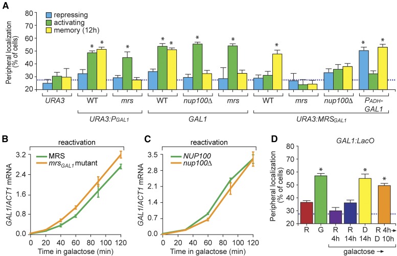
Memory Recruitment Sequence (MRS)GAL1-dependent peripheral localization of GAL1 during memory requires growth in glucose and Tup1. (A) Peripheral localization of URA3, GAL1, URA3:PGAL1, or URA3:MRSGAL1 was quantified under repressing (glucose), activating (galactose), and memory (galactose → glucose, 12 hr) conditions in wild-type (WT) or nup100∆ cells using immunofluorescence or live cell microscopy. The full-length GAL1 promoter (PGAL1, 667 bp) or the 63-bp MRSGAL1 were inserted at URA3 along with a LacO array as described (Egecioglu et al. 2014). The mrs mutation is shown in Figure S2B in File S1. (B and C) Cells were grown in galactose overnight, shifted to glucose for 12 hr, and then shifted to galactose (reactivation) to assay GAL1 expression using RT-quantitative PCR in WT, mrsGAL1 (B), and nup100∆ (C) mutant cells. (D) Peripheral localization of GAL1 in cells grown in raffinose (R), galactose (G), and upon shift from galactose to: raffinose for 4 hr (R 4 hr), raffinose for 14 hr (R 14 hr), glucose for 14 hr (D 14 hr), or raffinose 4 hr followed by glucose 10 hr (R 4 hr → D 10 hr). The hatched line represents the level of colocalization with the nuclear envelope predicted by chance (A and D). Error bars represent SEM for at least three biological replicates. * P ≤ 0.05 (Student’s t-test) relative to the repressing condition.

