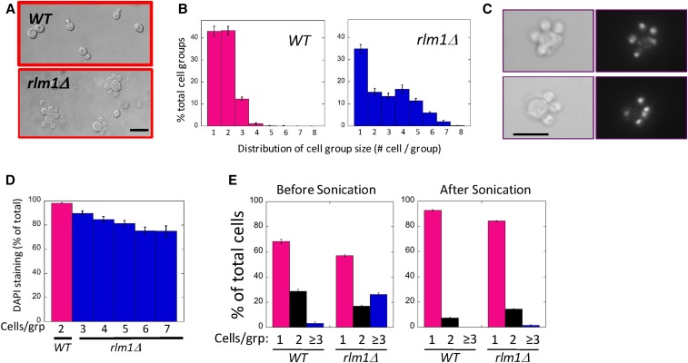
Figure 1.
The rlm1Δ mutant forms multi-budded cells. Wild-type (WT) (SH3881) and rlm1Δ (SH4708) spot colonies grown on LA medium for 24 hr, then resuspended and examined by microscope. (A) Representative wild-type (top) and rlm1Δ (bottom) cell groups visualized using Nomarski optics. Bar, 10 μm. (B) Distribution of the percentage of the total cell groups that contain indicated number of cells per group for both wild type (left) and rlm1Δ (right). n = 3. (C) Two examples of rlm1Δ satellite-cell groups stained with DAPI and visualized by bright-field (left) and fluorescent (right) microscopy. Bar, 10 μm. (D) Percentage of daughter cells that display nuclear DAPI staining for the wild type (magenta, n = 4) or rlm1Δ mutant (blue, n = 3) with the indicated number of cells / group. Wild-type groups containing more than two cells and rlm1Δ groups containing less than three cells are not shown. (E) Percentage of groups with one, two, or three or more cells for wild type or rlm1Δ before sonication (left panel) or after sonication (right panel), n = 3.

