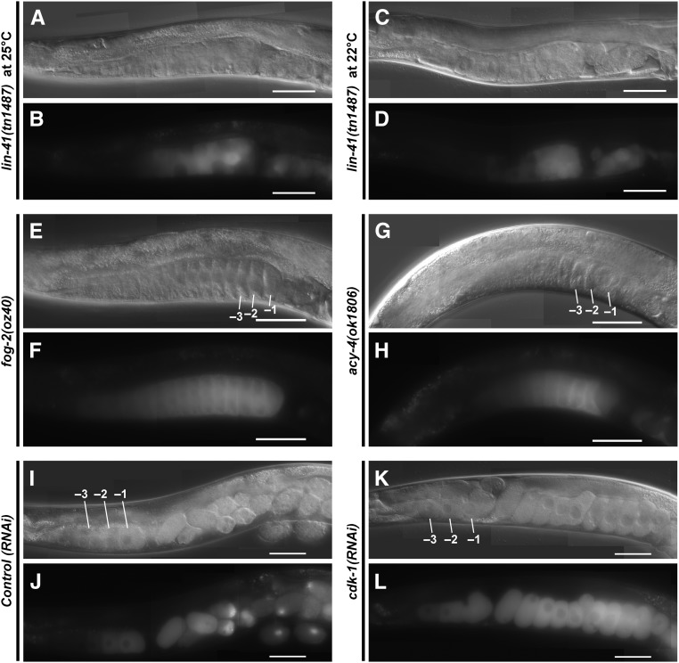
Figure 12.
Oocyte M-phase entry is not required for expression of GFP::3xFLAG::MEG-1. The expression of GFP::3xFLAG::MEG-1 was examined by fluorescence and DIC microscopy in (A–H) mutant backgrounds or (I–L) after RNAi treatments as indicated. GFP::3xFLAG::MEG-1 expression is observed in small abnormal oocytes in the lin-41(tn1487ts) mutant background (strain DG4274) at both (A and B) 25° and (C and D) 22°. A spatial expansion of GFP::3xFLAG::MEG-1 expression is observed in proximal oocytes in unmated fog-2(oz40) females [(E and F); strain DG4251] and acy-4(ok1806) hermaphrodites [(G and H); from strain DG4260]. (K and L) GFP::3xFLAG::MEG-1 expression is observed after cdk-1(RNAi). Bar, 50 μm.

