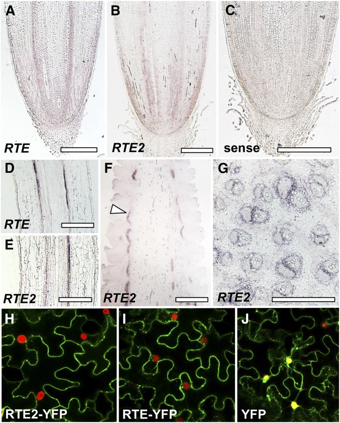
Figure 2.
Expression and subcellular localization analysis of RTE and RTE2. In situ hybridizations of longitudinal sections of seedling roots showing RTE and RTE2 expression in (A and B) root tips and (D and E) vasculature. (C) RTE2 sense control. (F) Longitudinal section of an immature ear showing RTE2 expression in vasculature (arrowhead). (G) Stem cross section. Bar, 500 µm. (H–J) Confocal images of tobacco epidermal cells expressing RTE2-YFP, RTE-YFP, YFP control, and the nuclear marker BAF1-mCHERRY (Gallavotti et al. 2011).

