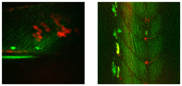
Fig. 7.
Image of a 5 days old zebrafish larva’s tail (left) and 2 days old larva’s muscle tissue (right). The FOV of both images is 300x300 µm2, the images are composed of 100 and 77 frames respectively. The green line structures are resulting from second harmonic radiation generated from collagen fibrils. The red structures are due to two-photon fluorescence from mCherry labelled cells. The (large) bright green/yellow structures visible are pigmented cells with stronger scattering.

