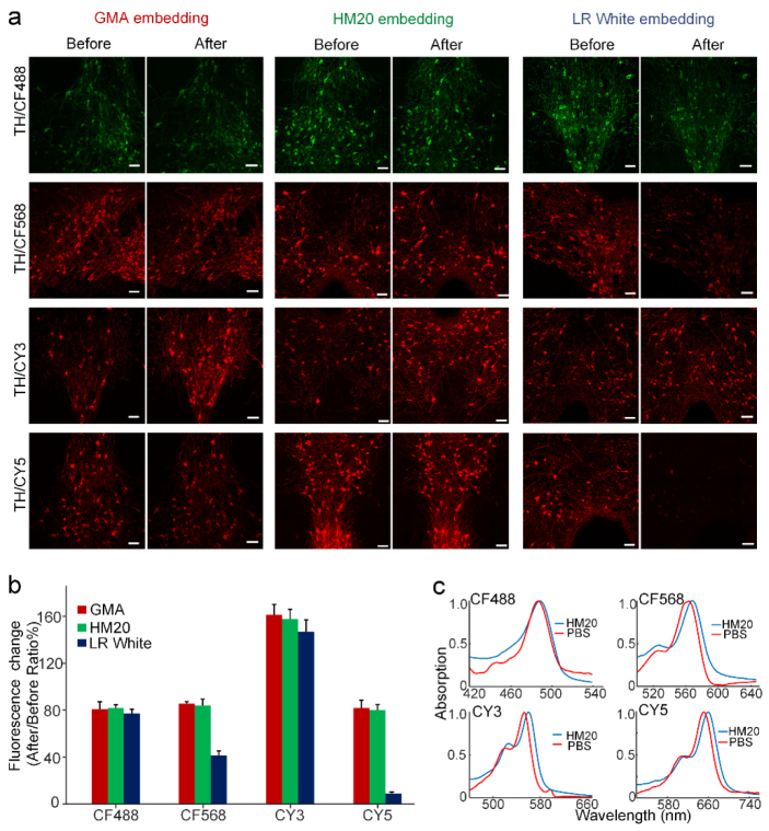
Fig. 1.
Immunofluorescence-labeled brain slices before and after resin embedding. (a) Images of labeled brain slices before and after GMA, Lowicryl HM20 or LR White resin embedding, respectively. All slices were immunolabeled by primary antibody (anti-TH), and were labeled with the various secondary antibodies conjugated with four fluorescent dyes: CF488, CF568, CY3, or CY5, respectively. All images were taken at a 0.42 × 0.42 × 1 μm3 voxel size on an LSM780 confocal microscope (ZEISS). (b) Fluorescent intensity change of immunostained brain slices after being embedding with different resins. We calculated the fluorescent intensity changes from labeled neuronal somas. The fluorescent intensities were compared before and after resin embedding using ImageJ software. The values in the bar graph are given as the means ± SD (n = 25 neurons from three slices for each independent sample). (c) Small shifts occurred in the absorption spectra of the dyes (CF488, CF568, CY3, and CY5) in Lowicryl HM20 resin polymer, compared to those in PBS buffer. Scale bar in (a): 50 μm.

