Fig. 2.
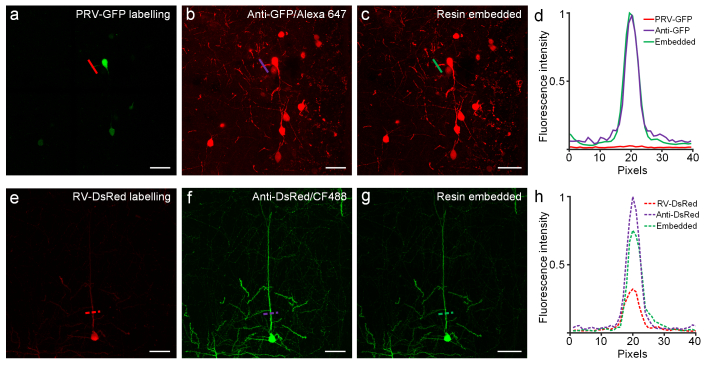
Preservation of immunofluorescence labeled fine details of neurite in resin-embedded brain tissue. (a) Fluorescence image of neurons labeled by PRV-GFP virus; slices were imaged after immunofluorescent labeling (b), and after resin embedding (c). (d) Fluorescent intensities of pixels crossed by red, purple, and green lines (shown in a, b, c, respectively) are plotted in the corresponding colors. (e) Morphology of neurons labeled by RV-DsRed virus before immunostaining, after immunofluorescent labeling (f), and after resin embedding (g). (h) Fluorescent intensities of pixels crossed by the red, purple, and green dashed lines (shown in e, f, and g, respectively) are plotted in the corresponding color. Images in panels (a-c) were recorded at a 0.42 × 0.42 × 1.00 μm3 voxel size using a confocal microscope (LSM780, ZEISS) with a 20 × 1.0 NA water objective. Images in panels (e-g) were recorded at a 0.21 × 0.21 × 1.00 μm3 voxel size using a confocal microscope (LSM780, ZEISS) with a 40 × 1.0 NA oil objective. All the images represent maximum intensity z-projections of 20-μm-thickness. Scale bar: (a-g) 50 μm.

