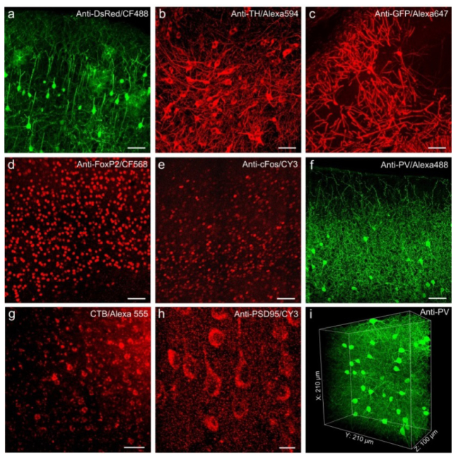
Fig. 3.
Resin embedding is compatible with various antibodies and fluorescent tracers staining in brain tissue. (a-i) Images of immune- and fluorescent tracer-labeled neurons in mouse brain tissue after Lowicryl HM20 resin embedding. (a) Immunolabeling of TdTomato-labeled mouse cortex from ChAT-cre; Rosa26lsl-tdTomato transgenic mice. (b) TH-immunolabeled thalamic neurons in C57 mouse brain tissue. (c) Immunolabeling of GFP-expressing hippocampal neurons from a Thy1-GFP-M transgenic mouse. (d-h) Mouse brain slices were immunolabeled by FoxP2 (d), cFos (e), parvalbumin (f), cholera toxin beta (g), and PSD-95 (h), respectively. Images in panel a-h were acquired on an LSM780 confocal microscope (ZEISS). (i) 3D volume image of immnofluorescent signals (Alexa 488) labeled by parvalbumin in mouse cortex, images were acquired by a two-photon microscope (LSM780). Scale bar: (a-g) 50 μm; (h) 10 μm.

