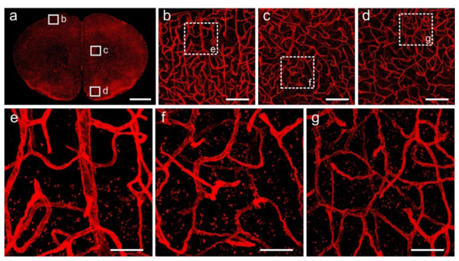
Fig. 4.
Fluorescent images of Lectin-DyLight 594 labeled vasculature in the brain tissue after Lowicryl HM20 resin embedding. (a) Maximum intensity projections of a 20-μm-thick coronal slice. (b-d) Corresponding magnification of regions indicated in (a). (e-g) High magnification images of the boxed regions in (b-d), respectively. All images were acquired using 20 × (NA = 1.0), at a 0.42 × 0.42 × 1.00 μm3 voxel size on a confocal microscope (LSM780, ZEISS). Scale bars: (a) 1 mm; (b-d) 100 μm; (e-g) 30 μm.

