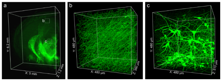
Fig. 5.
Imaging large volume immunolabeled mouse brain tissue after Lowicryl HM20 resin embedding. (a) 3D presentation of the TH immunolabeled mouse brain block. Enlargements of the fine structures of TH-positive axonal fibers in the cortex (b) and TH-positive soma located in the thalamus (c). Images were acquired by successive high-resolution stage-scanning microscopy at a 0.16 × 0.16 × 1.00 μm3 voxel size. (a) 6200 × 5000 × 1200 μm3; (b) 480 × 480 × 380 μm3; (c) 480 × 480 × 510 μm3.

