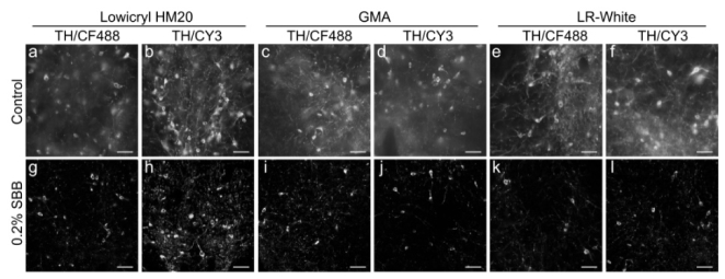
Fig. 6.
Effects of SBB on background fluorescence in the resin embedded brain tissue. Images obtained from immunostained brain tissue that were embedded in resin (HM20, GMA and LR white) with (g-l) and without 0.2% SBB (control, a-f), respectively. Mouse brain tissue was immunostained with anti-tyrosine hydroxylase primary antibody and CF488 or CY3-conjugated secondary antibodies. All images were recorded at 0.17 × 0.17 μm/pixel on the same wide field microscope (Nikon Ni-E) at room temperature. Scale bar: (a-l): 50 μm.

